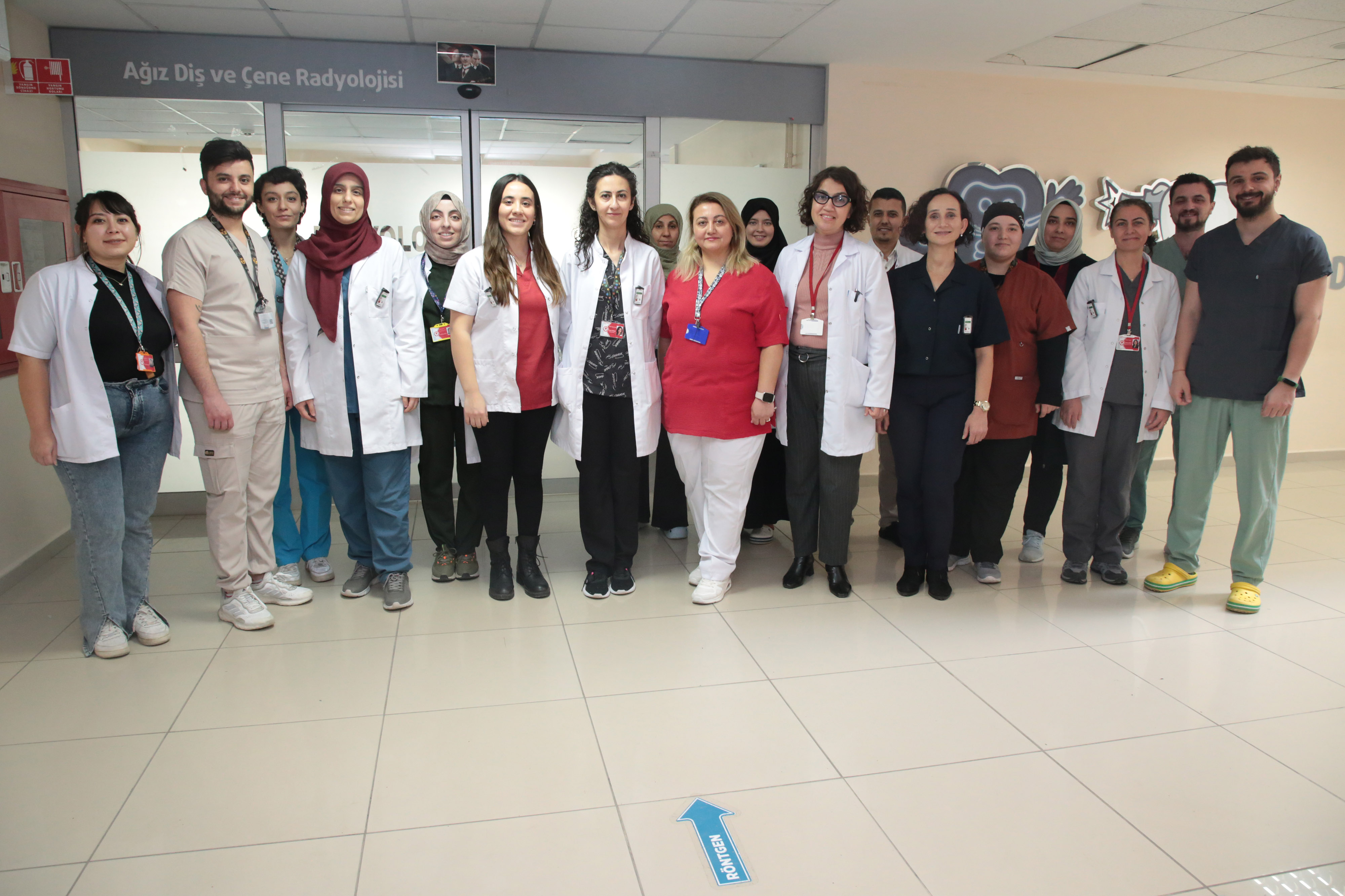
Oral and Maxillofacial Radiology
|
Chair
Academic Staff Assoc. Prof. Dr. Esin BOZDEMİR Lecturer Hasibe TAŞKIN
Communication 0246 211 32 54 |
HISTORY The Department of Oral and Maxillofacial Radiology is an academic division where oral and maxillofacial tissues and organs are systematically examined, all physiological and pathological changes of these structures are interpreted clinically and radiographically, and treatments to be performed are planned. Our department has trained a lot of Oral and Maxillofacial Radiology specialists so far.
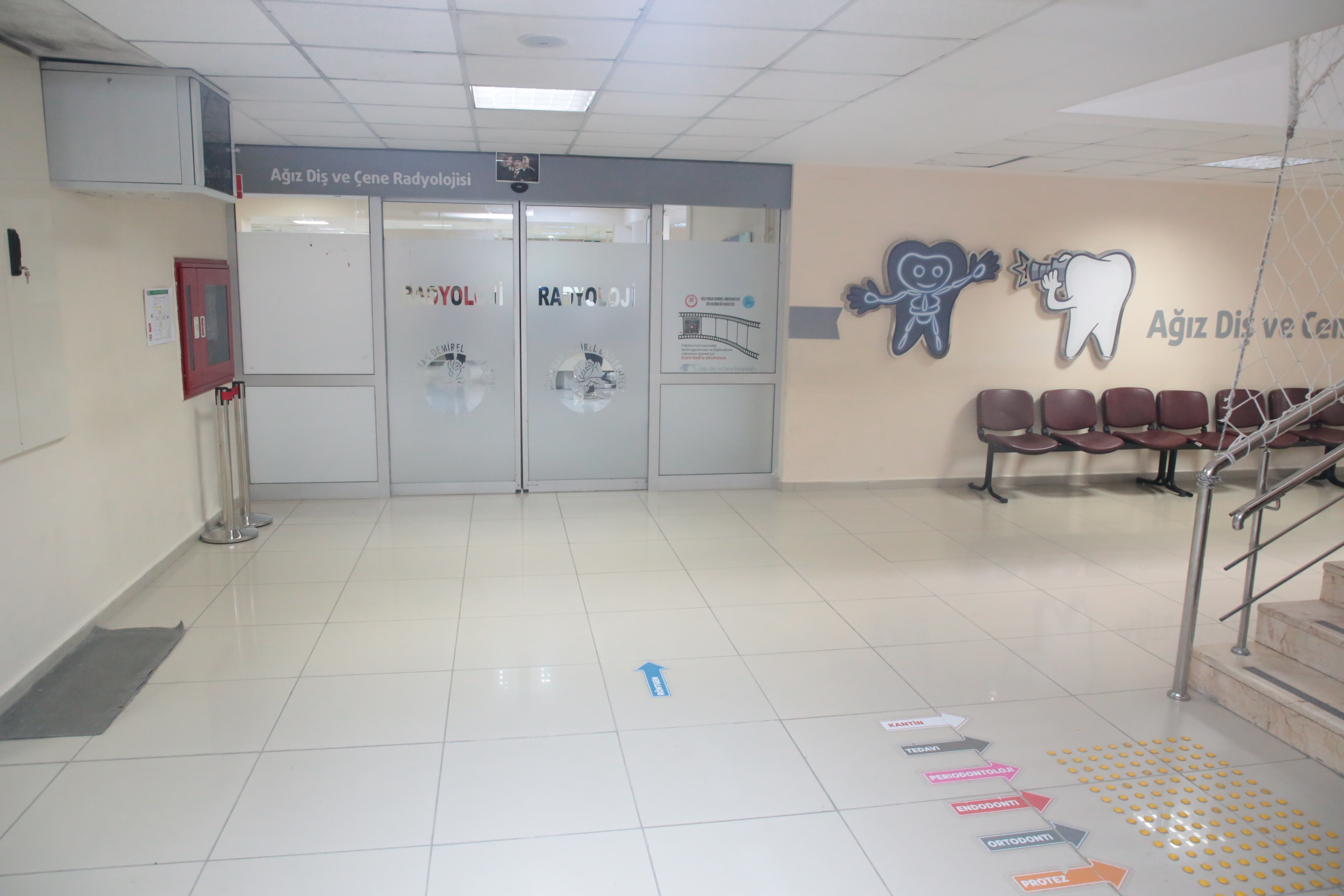
LOCATION and CAPACITY Our department continues its studies in two separate clinics, Oral Diagnosis and Radiology. In the Oral Diagnosis Clinic, the first examinations of the patients who apply to our Faculty are carried out with 11 units. After the examination in the Oral Diagnosis Clinic, intraoral or extraoral radiographs of the patient, which are deemed necessary for diagnosis, are taken in the Radiology Clinic. In the Radiology Clinic, 2 intraoral X-ray equipment, 1 conventional panoramic-cephalometric X-ray device, 2 digital panoramic-cephalometric radiographs are taken. X-ray device and 1 Cone beam computed tomography device are used.
RESEARCH ASSİSTANT TRAINING PROCESS The training period for research assistants (specialty students) is 3 years. During this training period, research assistants receive theoretical and clinical training, and make seminars, articles and case presentations. They rotate for 2 months in the Faculty of Medicine, Department of Radiology, and 1 month in the Department of Oral and Maxillofacial Surgery. At the end of the education period, research assistants who prepare a graduation thesis and take the final exam graduate with the title of specialist if they are successful.
DEPARTMENT OF STUDY AREAS Our Department is a basic academic unit in dentistry with its studies carried out in parallel with technological developments with new imaging devices developed day by day. The fields of study of our department are listed below: -Tooth and gum diseases - Jaw bones and temporomandibular joint - Soft tissues of the head and neck region - Oral diseases associated with systemic diseases - Physics and biology of radiation - Digital radiography OPPORTUNITIES - Oral Diagnosis Clinic - Radiology Clinic
TREATMENTS IN THE DEPARTMENT In the Oral and Maxillofacial Radiology Department, diseases in the mouth, teeth and jaw area are clinically and radiographically examined, diagnosed and the treatments to be performed are determined. Our department continues its studies in two separate clinics, Oral Diagnosis and Radiology. In the Oral Diagnosis Clinic, the first examinations of the patients who apply to our faculty are performed. Accurate diagnosis is essential for the patient to be treated appropriately and adequately. For this reason, all head-neck region and intraoral examinations are performed in the Oral Diagnosis Clinic, as well as examining the main reasons for patients' admission. Apart from dental caries and gingival diseases in the mouth, the diseases of the jaw bones and joints, salivary glands, tongue, floor of the mouth, lips and all soft tissues in the mouth are in the field of interest of our Department. Oral health cannot be considered separately from general body health. Many systemic diseases also show important symptoms in the mouth, and the symptoms of some of these diseases are first seen in the mouth. The relationship between oral diseases and systemic diseases is also examined in our department, and in this context, diagnosis and treatment planning of oral diseases is carried out. Due to some diseases and drugs used, precautions may need to be taken before dental treatments. For this reason, information is obtained about the general health of the patient at the time of application and in the past, as well as the drugs used. When deemed necessary, the patient is sent to the relevant branches of medicine for examination, and treatment planning is made in line with the reports sent. After the examination in the Oral Diagnosis Clinic, intraoral or extraoral radiographs of the patient are taken in the Radiology Clinic, which are deemed necessary for diagnosis. The disease is diagnosed in the light of clinical and radiological examinations and the patient is directed to the clinics where the necessary treatment will be applied. X-ray examination not only supports the findings of the oral examination, but also provides the diagnosis of diseases, cysts and tumors in the jaw bones that do not give any symptoms in the mouth. Today, imaging methods such as Magnetic Resonance Imaging, Ultrasonography, as well as dental and maxillofacial X-rays have become more and more used in dental radiology. For such radiological examinations, the patient is directed to medical radiology clinics. In our clinic, clinical and radiological evaluations of an average of 100-140 patients per day are performed.
MOST ASKED QUESTIONS AND ANSWERS BY PATIENTS Question 1: Is the first examination mandatory in dentistry? Existing dental diseases in patients and their treatment vary, and many patients may need more than one treatment. Various examinations are required to establish the diagnosis, which is the basis for the correct planning of these treatments. Pre-treatment examinations, requests and treatment plan are made during the first examination. Therefore, the first examination is mandatory for more comfortable and shorter treatment processes.
Question 2: Do I need to make an appointment to be examined? In order to be examined in our clinic, without the need for an appointment process, examinations are made and films are taken on the day of application during working hours. Patients are referred to other clinics for treatments.
Question 3: What are the documents that patients should bring with them when they come to the faculty for examination? Patients are required to bring their old patient cards, if any, and their previous films with them. In this way, re-filming will be prevented. If necessary, X-rays can be repeated by physicians. In addition, patients are required to bring their recent tests and results, as well as the medications they use. In this way, the physician will have information about the systemic condition of the person.
Question 4: Why are detailed questions asked about my health apart from my teeth during the examination? There may be a disease and a drug used during dental treatment that will affect the applications. In addition, some conditions in the mouth may be associated with systemic diseases. If correct and complete information is not given to the physician, life-threatening situations may be encountered during dental treatment or the factors of oral diseases may not be determined.
Question 5: Why is a dental X-ray taken? The purpose of X-ray is to diagnose pathologies in teeth, bones and soft tissue that cannot be seen during the examination. X-rays should be taken at the beginning of the treatment, during the treatment and after the treatment for control purposes. If the X-ray is not taken when necessary, the pathology and its cause cannot be determined correctly and a treatment plan cannot be made.
Question 6: Is dental X-ray harmful? In general, the radiation used in X-rays is harmful to living tissue. However, the radiation emitted from dental X-rays taken with modern devices and appropriate techniques is very low. In our clinic, service is provided by using modern devices, taking necessary precautions and applying appropriate techniques, and there is no medical problem.
Question 7: Is there any need for preparation before X-ray? Before X-ray, the patient should remove all metal items (earrings, buckles, necklaces, pins, glasses, piercings, removable prosthesis-dentures, hearing aids, etc.) in the head and neck area.
Question 8: Can pregnant patients take a X-ray? Radiological examination is not recommended during pregnancy, except in emergencies. In case of emergency, a minimum number of films can be taken after the necessary precautions are taken. The pregnant or suspected person must inform the physician of this situation.
Question 9: In which situations is computed tomography necessary? With cone-beam computed tomography available in our clinic, implant planning, examination of impacted teeth, evaluation of cysts and tumors in the head and neck region, detection of tooth and jaw traumas, diagnosis of degenerative jaw joint diseases and imaging of the sinuses are performed. The biggest advantage of this tomography compared to medical tomography is that the radiation dose received by the patient is much lower.
Informed Consent Forms |